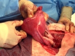
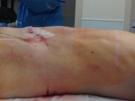
Note: this page contains actual photos of surgical procedures which some readers may find graphic
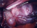
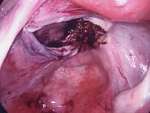
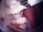
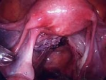
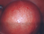
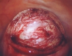
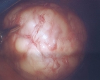
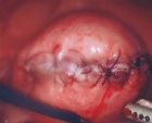
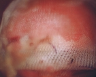
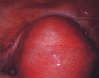
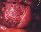
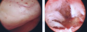
These photos show a variety of fibroid surgeries including abdominal myomectomy for very many or very large fibroids, laparoscopic myomectomy for fewer or not very large fibroids and hysteroscopic myomectomy for submucous fibroids.
Dr. Parker is a frequent lecturer and teacher of advanced surgical techniques both in the United States and abroad. He has published over 40 articles on his research in the areas of laparoscopic myomectomy, abdominal myomectomy, laparoscopic hysterectomy, and the care and treatment of women with fibroids.
While every fibroid surgery is slightly different, the goals are the same – remove all fibroids with minimal blood loss, perfectly reconstruct the uterus, and use an adhesion barrier to decrease the risk of scar tissue formation.
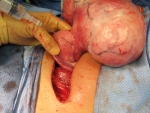
Abdominal Myomectomy, Patient A
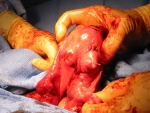
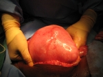
This woman had a very large pedunculated fibroid attached to her uterus. I injected a substance to decrease bleeding, removed the fibroid and reconstructed the uterus to essentially normal. (Click pictures to enlarge)
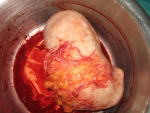
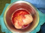
Abdominal Myomectomy, Patient B
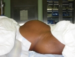
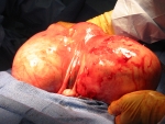
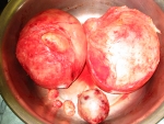
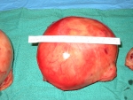
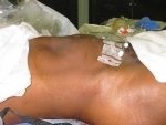
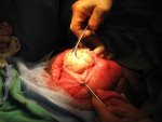
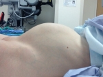
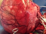
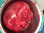
This woman had small fibroids diagnosed when she was 20 years old, but had been told by 10 different doctors that the only treatment was hysterectomy. So, she stopped going to doctors. I met her when she was 39 years old, and her fibroid uterus was the size of a 9 month pregnancy. I performed an abdominal myomectomy and removed two fibroids that were each 15 cm (7 inches) and four smaller fibroids. We used the cell saver, but there was very little blood loss and not enough to give her back at the end of surgery. I recently saw her, now a year after surgery, and she has a flat abdomen, her uterus is entirely normal size, and she feels great. This surgery changed her life and it was very rewarding for me to be able to help her. (Click pictures to enlarge)
Abdominal Myomectomy for Multiple Fibroids, Patient C
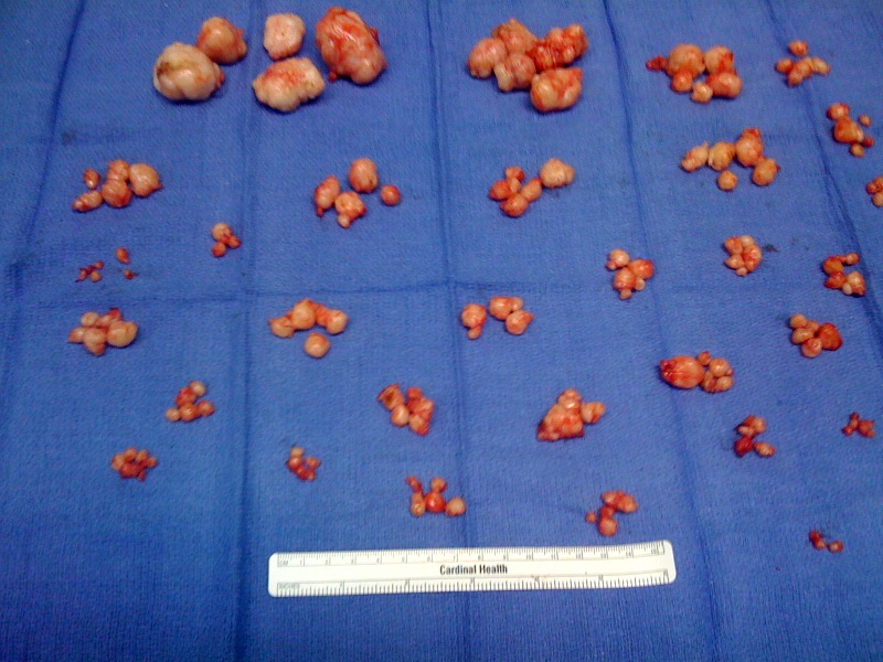
This woman had 150 small fibroids removed. With careful surgical technique, blood loss was minimal and she did very well. About 20 fibroids were removed from inside the uterine cavity and a hysteroscopy performed in my office 6 weeks after surgery showed a normal uterine cavity. Since the uterine lining cells re-grow every month, the lining has excellent healing capabilities.
Abdominal Myomectomy, Patient D
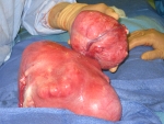
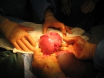
A very large uterus with many intramural fibroids and one large pedunculated fibroid. After the surgery, the uterus is still swollen, but returned to normal size 8 weeks after surgery. The uterus has a remarkable ability to heal. This is readily apparent after childbirth, when a nine month size pregnant uterus returns to normal size in about 10 weeks. (Click pictures to enlarge)
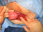
Abdominal Myomectomy, Patient E
This woman had a large uterus with many fibroids and had been told that myomectomy was not possible. Surgery went without problems.
Laparoscopic Myomectomy, Patient F
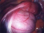
Laparoscopic myomectomy can be performed on large fibroids. This fibroid is near the ureter, but was carefully peeled away. The fibroid was morcellated to remove it from the pelvic cavity. The uterus is essentially normal at the end of the procedure. (Click pictures to enlarge)
Laparoscopic Myomectomy, Patient G
This woman was 35 and had a 7cm fibroid causing her abdominal pressure and urinary frequency. The fibroid was removed laparoscopically as an outpatient and she returned to work in 7 days.
Adenomyosis, Patient H
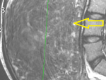
This woman had severe menstrual cramps and heavy bleeding with her periods. Her uterus was enlarged to a 3 month pregnancy size and an MRI revealed severe adenomyosis. Unfortunately, hysterectomy is the only consistently successful treatment for adenomyosis (UAE works about 50% of the time). She had a laparoscopic supracervical hysterectomy and is now pain-free.
Hysteroscopic Myomectomy, Patient I
This 35 year old woman had very heavy menstrual bleeding, requiring her to change menstrual pads every hour for 2 full days a month and causing her to be anemic. A hysteroscopy performed in the office showed a fibroid in the uterine cavity. This fibroid was removed hysteroscopically (an outpatient hospital procedure) and she was back to work in 3 days. She is now 1 year after surgery and has entirely normal periods and plans to get pregnant soon.
Abdominal Myomectomy, Patient J
A 34 year-old woman who has not had children, and who had been told by multiple gynecologists that a hysterectomy was her only option. So, she stopped seeing gynecologists
William H. Parker, MD
Clinical Professor, Reproductive Medicine, UC San Diego School of Medicine
Page last updated: January, 2018


