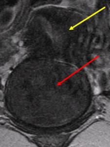
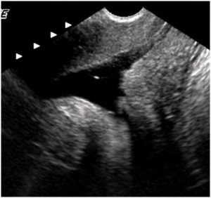
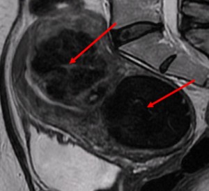
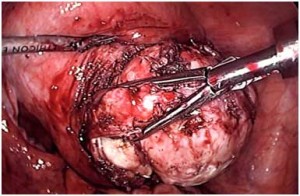
MRI is the most accurate diagnostic test we have to tell how many fibroids you have, the precise sizes of the fibroids and the position of each fibroid within the uterine wall (submucous, intramural, subserous). This information can help a knowledgeable gynecologist help you decide what treatments might be appropriate. The best gynecologists will understand that many treatment options exist: watchful waiting; medical treatment for heavy bleeding with medication or the Mirena IUD; hysteroscopic, laparoscopic or abdominal myomectomy; uterine artery embolization; focused ultrasound;
How Does MRI Work?
Your body is made up mostly of water. The MRI scanner contains a very powerful magnet which changes the position of the protons in the water molecules. When the protons return to their normal positions they give off a radio wave that is translated by the MRI computer into an image. Tissues that have a lot of water look white on certain MRI images.
Patient Stories
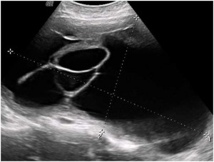
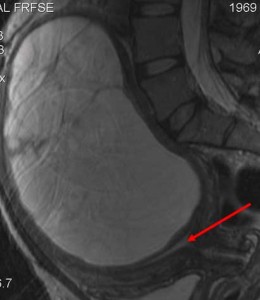
A 40 year-old woman with one year of very heavy bleeding resulting in severe anemia was seen elsewhere and an ultrasound diagnosed a large ovarian cyst. The gynecologist thought the patient might have ovarian cancer and suggested immediate surgery. A large abdominal incision was made, but the ovaries looked entirely normal. The uterus, however, was enlarged, but the doctor didn’t know why and closed the incision without any surgery accomplished.
When I saw her, I ordered an MRI which showed the cyst was inside the uterine wall – not the ovary. This picture was classic for cystic degeneration of a fibroid and I was able to remove just the fibroid and preserve her uterus, per her wishes.
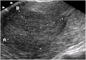
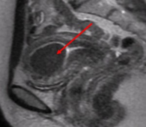
A 40 year-old woman with very heavy bleeding was treated unsuccessfully with birth control pills and then her gynecologist recommended an abdominal hysterectomy. Her ultrasound showed a fibroid near the uterine cavity, but the MRI showed the fibroid clearly inside the cavity. As a result, I was able to remove the fibroid using a hysteroscope, as an outpatient. She was back to normal activity in 2 days, rather than the 6 weeks need for recovery from the recommended treatment.
A 48 year-old woman with heavy bleeding. An ultrasound diagnosis was a fibroid behind the uterus attached by a stalk. One gynecologist suggested an abdominal hysterectomy, another suggested uterine artery embolization. The MRI shows a fibroid coming from inside the cervix and bulging into the vagina. Based on this information, I was able to remove the fibroid through the vagina, without any incisions and as an outpatient, with a 2 day recovery.
A 35 year-old woman with pelvic discomfort and a 3½ month size uterus was treated by embolization without any improvement. The saline-infusion ultrasound showed an irregular uterine cavity and two fibroids. Her gynecologist then recommended an abdominal hysterectomy.
The MRI showed two fibroids behind the uterus and laparoscopic myomectomy was clearly possible.
William H. Parker, MD
Clinical Professor, Reproductive Medicine, UC San Diego School of Medicine
Page last updated: January, 2018


