Topics of Interest

Dr. Parker's Fibroids Blog

Finally: Prevention of Ovarian Cancer!

Ovarian cancer is a terrible disease, almost impossible to find early before it has spread and very hard to cure despite the best efforts over the past 40 years. As a result, it had been common practice to remove ovaries when a hysterectomy was performed in order to prevent the future development of ovarian cancer. However, it has also been known for 40 years that the ovaries continue to produce hormones after menopause (and forever), although in smaller amounts. I have been the principal author of three studies that show that women who have their ovaries removed had a higher risk of death from heart attack, lung cancer and colon cancer (abstracts below). As a result, I have been encouraging women to keep their ovaries if they had to have a hysterectomy, unless they were at known high-risk of ovarian cancer due to the BRCA genes or a strong family history.

Surprisingly, in the past few years, it has become clear that the most aggressive types of “ovarian” cancer don’t start in the ovaries at all. Most “ovarian cancer” actually starts in the fallopian tube and some of these cancers start in the uterine lining cells that are shed and pass out the ends of the tubes during a normal menstrual period. So, after all of these years, most “ovarian” cancer is, in fact, fallopian tube or uterine lining cell cancer. Since the fallopian tubes are open at the end, this explains why the cancer spreads so quickly and easily. It also explains why screening for early ovarian cancer by looking at the ovaries with ultrasound has been such a dismal failure.

After this recent discovery of “fallopian tube cancer”, a number of hospitals in Canada and Europe began recommending that women have their fallopian tubes removed, rather than their ovaries, when they were having hysterectomies. I have been concerned though, since the tube and ovary share some common blood vessels, that removal of the tube might reduce blood flow to the ovaries and diminish hormone production. If this were the case, women still might end up with the long-term health consequences that we found in the studies we did (abstracts below).

The good news is that a new study, just published in Gynecologic Oncology, shows that ovarian function as measured by a number of blood and ultrasound tests (AMH, FSH, ovarian volume and antral follicle counts) remains normal following removal of the tubes. As a result, it appears to be safe to remove the tubes. I am, therefore, going to start discussing with all of my patients who do not want any more children, the option of removing the fallopian tubes at the time of pelvic surgery for any reason: myomectomy, ovarian cyst removal, hysterectomy, etc.

My thought, which I am going to write in a letter to the editor of Gynecologic Oncology, is that gynecologists should also start teaching general surgeons how to perform removal of the tubes and that any woman (not interested in childbearing) who is having abdominal surgery should consider removal of the fallopian tubes at the same time.

Ovarian cancer has been a frustrating story, but now it likely has a happy ending – hopefully, the prevention of most tubal (“ovarian”) cancers that have caused so much ill health, death and fear.

Our research, showing the benefit of keeping your ovaries, will end up as part of the larger story; since ovarian cancer was never ovarian, the ovaries should never have been removed all along!

References

Prophylactic Salpingectomy in Premenopausal Low-risk Women for Ovarian Cancer: Primum non nocere.
Morelli M, Venturella R, Mocciaro R, Di Cello A, Rania E, Lico D, D’Alessandro P, Zullo F. Gynecologic Oncology. 2013;129:448-51.

Long-Term Mortality Associated With Oophorectomy Compared With Ovarian Conservation in the Nurses’ Health Study.
Parker WH, Feskanich D, Broder MS, Chang E, Shoupe D, Farquhar CM, Berek JS, Manson JE.
Obstetrics and Gynecology. 2013;121:709-716.

OBJECTIVE: To report long-term mortality after oophorectomy or ovarian conservation at the time of hysterectomy in subgroups of women based on age at the time of surgery, use of estrogen therapy, presence of risk factors for coronary heart disease, and length of follow-up.

METHODS: This was a prospective cohort study of 30,117 Nurses’ Health Study participants undergoing hysterectomy for benign disease. Multivariable adjusted hazard ratios for death from coronary heart disease, stroke, breast cancer, epithelial ovarian cancer, lung cancer, colorectal cancer, total cancer, and all causes were determined comparing bilateral oophorectomy (n=16,914) with ovarian conservation (n=13,203).

RESULTS: Over 28 years of follow-up, 16.8% of women with hysterectomy and bilateral oophorectomy died from all causes compared with 13.3% of women who had ovarian conservation (hazard ratio 1.13, 95% confidence interval 1.06-1.21). Oophorectomy was associated with a lower risk of death from ovarian cancer (four women with oophorectomy compared with 44 women with ovarian conservation) and, before age 47.5 years, a lower risk of death from breast cancer. However, at no age was oophorectomy associated with a lower risk of other cause-specific or all-cause mortality. For women younger than 50 years at the time of hysterectomy, bilateral oophorectomy was associated with significantly increased mortality in women who had never used estrogen therapy but not in past and current users: assuming a 35-year lifespan after oophorectomy: number needed to harm for all-cause death=8, coronary heart disease death=33, and lung cancer death=50.

CONCLUSIONS: Bilateral oophorectomy is associated with increased mortality in women aged younger than 50 years who never used estrogen therapy and at no age is oophorectomy associated with increased survival.

LEVEL OF EVIDENCE: I

Ovarian Conservation at the Time of Hysterectomy and Long-term Health Outcomes in the Nurses’ Health Study.
Parker WH, Broder MS, Chang E, Feskanich D, Farquhar C, Liu Z, Shoupe D, Berek JS, Hankinson S, Manson JE.
Obstetrics and Gynecology. 2009;113:1027-37.

OBJECTIVE: To report long-term health outcomes and mortality after oophorectomy or ovarian conservation.

METHODS: We conducted a prospective, observational study of 29,380 women participants of the Nurses’ Health Study who had a hysterectomy for benign disease; 16,345 (55.6%) had hysterectomy with bilateral oophorectomy, and 13,035 (44.4%) had hysterectomy with ovarian conservation. We evaluated incident events or death due to coronary heart disease (CHD), stroke, breast cancer, ovarian cancer, lung cancer, colorectal cancer, total cancers, hip fracture, pulmonary embolus, and death from all causes.

RESULTS: Over 24 years of follow-up, for women with hysterectomy and bilateral oophorectomy compared with ovarian conservation, the multivariable hazard ratios (HRs) were 1.12 (95% confidence interval [CI] 1.03-1.21) for total mortality, 1.17 (95% CI 1.02-1.35) for fatal plus nonfatal CHD, and 1.14 (95% CI 0.98-1.33) for stroke. Although the risks of breast (HR 0.75, 95% CI 0.68-0.84), ovarian (HR 0.04, 95% CI 0.01-0.09, number needed to treat=220), and total cancers (HR 0.90, 95% CI 0.84-0.96) decreased after oophorectomy, lung cancer incidence (HR=1.26, 95% CI 1.02-1.56, number needed to harm=190), and total cancer mortality (HR=1.17, 95% CI 1.04-1.32) increased. For those never having used estrogen therapy, bilateral oophorectomy before age 50 years was associated with an increased risk of all-cause mortality, CHD, and stroke. With an approximate 35-year life span after surgery, one additional death would be expected for every nine oophorectomies performed.

CONCLUSION: Compared with ovarian conservation, bilateral oophorectomy at the time of hysterectomy for benign disease is associated with a decreased risk of breast and ovarian cancer but an increased risk of all-cause mortality, fatal and nonfatal coronary heart disease, and lung cancer. In no analysis or age group was oophorectomy associated with increased survival.

View all blog archives

Dr. Parker featured on NBC4 Los Angeles

Dr. Parker was interviewed for NBC4 Los Angeles regarding his latest study on removing a woman’s ovaries during hysterectomy.

Removing Ovaries During Hysterectomy May Increase Health Risks: Study

A new study finds that hysterectomies may cause more harm than good. The study revealed that even after a woman’s reproductive years are over, her ovaries are protecting other parts of the body. Dr. Bruce Hensel reports for the NBC4 News at 5 p.m. on June 13, 2013.

View all blog archives

Degenerating Fibroid

Hello Dr. Parker,

I have read your website https://www.fibroidsecondopinion.com/ and it has provided me with a wealth of information. I hope you don’t mind me asking you a question in regards to degenerating fibroids.  Do degenerated fibroids need to be removed? Is there harm to having necrotic tissue in the body? Two began to degenerate while I was pregnant last year. Now one shows central areas of necrotic tissue.

Thanks,

Natalie


Hi Natalie,

There is no danger to having degenerated fibroids in your body.  Sometimes the degeneration can cause pain or low grade fever, but usually nothing more than that.  The degenerated fibroid is a bit like the inside of a tree – not dead or decomposed, but not as alive as normal tissue.  Sometimes some of the degenerated tissue is reabsorbed by the body, but often it stays there and causes no problems.

The following is excerpted from: https://www.fibroidsecondopinion.com/fibroid-symptoms/

Fibroids are living tissue, and need blood and oxygen to survive. If a fibroid grows quickly, blood vessels feeding the fibroid may not be able to grow fast enough to supply the new tissue with enough blood and oxygen. If this happens, the fibroid undergoes a process called degeneration, or cell death. As the cells in the fibroid die, chemical substances are released that cause pain and swelling in the uterus. This pain may be severe but is not usually associated with any serious problems. If these chemical substances from a degenerating fibroid reach the bloodstream, they may cause a low fever. As some of the fibroid dies, the blood supply to the rest of the fibroid will be enough to keep it alive and healthy. At this point, the pain will go away. This process may take a few weeks. When pain develops in a woman with fibroids, examination by a physician is important to help figure out the source of the problem.

If you have a degenerating fibroid, a heating pad on your abdomen will be comforting, and pain medication should provide relief for a few days or weeks until the pain begins to subside. In rare instances, a fibroid on a stalk (pedunculated fibroid) can twist around on the stalk so that no blood can get through the stalk to the fibroid. If that happens, the entire fibroid begins to die, and the pain becomes very severe and surgery is usually necessary to remove the dying fibroid.

I hope this is helpful,
Bill Parker, MD


View all blog archives

Dr. William Parker joins UCLA faculty full-time

Dr. William Parker is pleased to announce his appointment as Director of Minimally Invasive Gynecologic Surgery for UCLA. While joining the faculty of UCLA full-time, Dr. Parker will continue to maintain his surgical practice at his current office location in Santa Monica.

This appointment enables Dr. Parker to do more teaching with the faculty and residents of UCLA. His focus will be to share his surgical expertise and special skills and to teach gynecology residents about the many alternatives to hysterectomy for women with fibroids, ovarian cysts and other benign conditions.

As a benefit to many patients, health insurance plans that include coverage with UCLA will now be accepted. Consultation appointments can be made, as they have always been, by calling (310) 451-8144.

View all blog archives

What Size are My Fibroids?

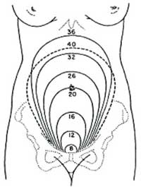
Gynecologists and ultrasound reports usually describe the size of fibroids using centimeters (cm), a measurement unfamiliar to most Americans.  Often, a comparison to fruits or common objects can help to understand the size.  We also compare the size to weeks or months of pregnancy, since many women are familiar with this measurement.  The following measurements may help:

Pea = 1 cm

Pea = 1 cm

Grape = 2 cm

Grape = 2 cm

Walnut = 3.5 cm

Walnut = 3.5 cm

Plum = 5 cm

Plum = 5 cmTennis ball = 6.5 cm

Orange = 6.6 cm

Orange = 6.6 cm

Baseball = 7.5 cm

Baseball = 7.5 cm

Grapefruit = 10 cm

Grapefruit = 10 cm

Cantaloupe = 12 cm

Cantaloupe = 12 cm

Honeydew melon = 16 cm

Honeydew melon = 16 cm

Uterine size in weeks of pregnancy

Uterine size in weeks of pregnancy


View all blog archives

Un-Watchful Waiting

Over the years, a number of women have come to my office with extreme symptoms due to uterine fibroids. Some of these women had fibroids that were very large (the size of a full-term pregnancy), some had severe bleeding that caused a very low blood count (hemoglobin of 4- 6, when 12 is normal), and some had fibroids that blocked the bladder and required a visit to the emergency room for a urinary catheter to be inserted.

All of these intelligent women were aware that they had fibroids. However, they had put off treatment after having been told by numerous doctors (as many as 7 in one woman’s case) that the only remedy was a hysterectomy. Some of the women had not yet had children and wanted a family. Many felt strongly about keeping the body parts they were born with, and others feared a hysterectomy. Since they didn’t want or medically need a hysterectomy, they just stopped seeing doctors.

However, these women tolerated terrible symptoms and allowed fibroids to control their lives. Some were unable to leave the house every month when the bleeding was heavy. Some were weak and tired and weren’t able to function at work or at home. Some reported having strangers asking them “when the baby is due”. Most were consumed with worry about their health and about what was going to happen to them.

My sense is that these women accommodated to their symptoms over time. “Well, I am bleeding heavily but not much heavier than last month.” “Well, the fibroids are larger but not that much larger than 6 months ago.” The problem is that “normal” had been forgotten, and slow growth or a slow increase in bleeding can be deceptive. Following a myomectomy, these women often say “I forgot how good it feels to have a fibroid-free life” or “I feel like a new person”.

However, treatment options may be limited when the fibroids are very large, or if a woman has a very low blood count. For instance, laparoscopic or robotic myomectomy is not possible if the uterus is so large that there is not enough room to place the instruments of the robot in the abdomen. Some interventional radiologists do not recommend uterine artery embolization for very large fibroids because they think the procedure is not likely to be successful. Severe anemia may require either iron infusions or even, rarely, a blood transfusion before anesthesia and surgery can proceed safely.

Watchful waiting is an excellent solution for many women with fibroids. Some fibroids don’t grow, and some even shrink before menopause. After menopause most fibroids will shrink and bleeding will stop. My advice though is to continue to search for a gynecologist who has experience with managing fibroids on a regular basis, who can help you with the “watching” and who is experienced at treatment options including myomectomy in case surgery becomes necessary.

In my practice, I like to see women with bothersome fibroids on a regular, usually every 3- 6 months, basis. On these visits, I review their symptoms and examine them. We monitor blood counts when bleeding gets heavier, and re-discuss treatment options if symptoms get worse, or if the patient has reached the end of her rope. So, watchful waiting is fine, but have someone help you “watch”.


View all blog archives

Adenomyosis and Infertility

Journal: Obstetrics and Gynecology International, 2012

Authors: Campo S, Campo V, Benagiano G.

Study from: Catholic University of Sacred Heart, Roma, Italy.

Problem: Adenomyosis is a benign condition where the uterine lining cells grow deep into the uterine muscle wall. For many years, adenomyosis has been suspected of causing infertility in women. In the past, the diagnosis of adenomyosis has only been possible based on a pathologist’ examining the uterine muscle and lining cells after a hysterectomy, so fertility could never be measured. Now MRI (and probably 3-D ultrasound) allows the diagnosis of adenomyosis without hysterectomy and, therefore, scientific investigation of this condition has increased dramatically.

Study: The authors reviewed the new research and they present multiple reasons why adenomyosis might cause infertility.

Results: The evidence that adenomyosis causes infertility includes:

  1. In women with adenomyosis, the muscles cells in the uterine wall swell and they function differently than normal uterine muscle cells.
  2. In women with adenomyosis, the lining cells of the uterus do not go through normal changes during the menstrual cycle.
  3. In women with adenomyosis, the uterine lining cells cause inflammation in the muscle wall which interferes with the movement of the fertilized egg inside the uterus.
  4. In women with adenomyosis, there are changes in the uterine lining cells that interfere with implantation of the fertilized embryo.
  5. Surgical removal of adenomyosis or medical treatment with GnRH agonists (Lupron) increases the chance of pregnancy.

Dr. Parker’s Comments: In order to prove that adenomyosis causes infertility, a large group of women would need to have an MRI before trying to get pregnant and the percent of women who get pregnant with adenomyosis would need to be compared with the percentage of women who do not have adenomyosis. Since that type of study has never been done, it is currently impossible to prove that adenomyosis can lead to infertility. However, now that women with adenomyosis can be diagnosed by MRI, careful examination of the cellular changes of the uterus suggests that there are many reasons why adenomyosis could cause infertility.

These new studies will also be very important in helping scientists figure out what type of treatments might cure adenomyosis, or better yet, prevent it from developing at all.


View all blog archives

Predictors of Successful Surgical Outcome in Laparoscopic Hysterectomy

Authors: Twijnstra AR, Blikkendaal MD, van Zwet EW, van Kesteren PJ, de Kroon CD, Jansen FW.

Journal: Obstetrics and Gynecology. 2012;119:700-8.

Study from: Leiden University Medical Center, the Netherlands.

Problem:  Can a successful surgery be predicted from the surgeon’s experience or other measures of his/her surgical skill.

Study: The outcomes of 1,534 laparoscopic hysterectomies performed by 79 surgeons were studied.

Results:  Once a surgeon performed 125 surgeries there was less risk of complications during surgery.  Other factors that increased the risks of complications were a larger uterus (large fibroids), a heavier patient and women with scar tissue from previous abdominal surgeries.  The authors also identified an individual “surgical skills factor” suggesting that some surgeons are better than others.

Authors’ Conclusions:  An increase in the surgeon’s experience predicted a successful outcome with respect to less bleeding and complications. However, there was a large variation in proficiency between individual surgeons. The fact that a surgeon has performed many laparoscopic hysterectomies did not necessarily guarantee a good surgical outcome.

Dr. Parker’s Comments: In an earlier post, I reviewed an article showing that surgeons who operated on a regular basis had better results than surgeons who operated infrequently.  The article reviewed here reinforces this idea and suggests that it takes about 125 procedures to become an expert surgeon. The study also suggests that despite performing 125 procedures, some surgeons will be better than others.

Over the 20 years I have been training residents and fellows, differences in surgical skills and judgment have been noticeable. Some gynecologists-in-training have it and some don’t.  In addition to learning the anatomy of the female pelvis and learning which instruments work best for each part of the procedure, skills such as hand-eye co-ordination, moving in a 3-dimensional space and an unhurried, thoughtful approach to surgical technique seem to be more natural talents that are hard to teach and may not improve even with practice.

As I stated in that earlier post, it is important to ask your surgeon how many procedures like yours they have performed.  But, it is also important to ask if they have had any complications and what were they.

View all blog archives

Recovery at Home After Major Gynecologic Surgery

Authors: Evenson M, Payne D, Nygaard I.

Source: Obstetrics and Gynecology, 2012;119:780-4

Study from: University of Utah School of Medicine

Problem: Women’s recovery and pain control during the first 6 weeks following gynecologic surgery has not been well studied.

Study: Women having either abdominal or vaginal surgery were asked to determine their pain and pain medication use after surgery.

Results:  80 women had vaginal surgery; some women had a hysterectomy and others had bladder repair surgery. 47 women had abdominal surgery; some women had a hysterectomy and others had removal of fibroids (myomectomy) or ovarian cysts.

On recovery day 3, inadequate pain control was reported by about 50% of women in both groups and at the end of one week 25% of women had inadequate pain control.  By day 14, most women who had vaginal surgery had minimal pain, but 20% of women still had pain after abdominal surgery.  Two weeks after discharge, narcotic pain medication was still being used by 10% of women after vaginal surgery and 32% of women after abdominal surgery.  By 6 weeks, half of the women in each group felt recovered and two-thirds felt “back to normal.”

Authors’ Conclusions: Pain control after hospital discharge is inadequate for many women after both vaginal and abdominal surgery. The time to full recovery is longer than 6 weeks for half of women.

Dr. Parker’s Comments: Recovery from surgery varies from woman to woman, but it usually takes a bit longer than either the doctor or patient hopes will be the case.  In the past 10 years, doctors have been better educated about helping patients manage pain, but some doctors still discourage use of pain medicines and many patients stop using pain medicine before they should.   Laparoscopic surgery has been proven to cause less pain and have a faster recovery than abdominal or vaginal surgery. But, even following laparoscopic surgery, patients often try to return to normal activity faster than they should.

I always tell patients that there is no advantage to being a martyr and that they will sleep better and feel better if they take some pain medication for the first few weeks after surgery. Also, patients are not aware of the level of fatigue that they will feel after surgery. And, the fatigue can last for weeks after the pain has gone away.  Fatigue is due to a number of things: the effects of anesthesia and narcotic pain medication; the stress of surgery on the body; tissue injury and release of proteins (cytokines) into the bloodstream that are sedating (nature’s way to force you to rest); and, injured tissues need for energy to heal, leaving little reserve for the rest of your body.

So, some form of pain medication allows you to rest more and sleep better.  Also understand that recovery and return of energy may take longer than you wish.

View all blog archives

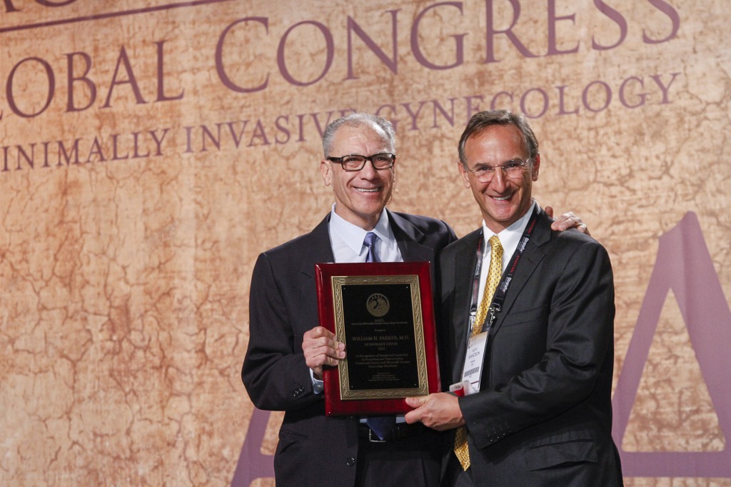
Dr. Parker Selected as Honorary Chair, Global Congress of Minimally Invasive Surgery – November, 2012


Dr. Bill Parker was selected as the Honorary Chair for the Global Congress of Minimally Invasive Surgery, attended by 2,000 gynecologists from more than 60 countries.

Dr. Parker’s Honorary Lecture, Everything You Learned in Your Residency Will Turn Out to be Wrong, called attention to the resistance to change in medicine despite rapidly-evolving science and the opportunity to share new information over the internet. He stressed that, for gynecologists, the misguided reliance on hysterectomy as often the first recommendation for gynecologic problems was based on old teaching. The hysterectomy recommendation often is easier, more profitable or more fun for the doctor, rather than what is right for the patient.

Dr. Parker also asked for the acronym Minimally Invasive Surgery (MIS) to be redefined as Minimally Invasive Solution (MIS), with diminished emphasis on surgery and increased emphasis on alternative methods. If surgery is necessary, the focus should be on uterine-conserving procedures – suggesting that even for large fibroids, women often prefer an abdominal myomectomy through a bikini incision instead of laparoscopic or robotic hysterectomy.

Dr. Parker encouraged new doctors to keep learning and to keep an open mind to new ideas. Although technology may bring some benefits, the simple ideas of close patient relationships, operating room patient-safety principles, and kindness should be encouraged above all. Dr. Parker ended the talk by asking doctors to have the intellectual humility to understand that new and better ideas will always on the way. The 2,000 attendees of the conference gave Dr. Parker a standing ovation.

Video of Dr. Parker’s lecture is below:


View all blog archives

Share/Save/Bookmark

Disclaimer: The ideas, procedures and suggestions contained on this web site are not intended as a substitute for consulting with your physician. All matters regarding your health require medical supervision.

Fibroid Doctor William H. Parker

Dr. William H. Parker is a board-certified Fellow in the American College of Obstetricians and Gynecologists. Dr. Parker is an internationally recognized expert in fibroid surgery and research. Based in San Diego, California, he is considered one of the best fibroid surgeons for abdominal and laparoscopic myomectomy in the United States and abroad. He has been chosen for Best Doctors in America and Top Doctors every year beginning in the late 90's.

Free Email Updates

Powered by Google FeedBurner : Receive free updates right to your inbox every time new content is added to this website.


Subscribe
Schedule an Appointment Today!
Read Dr. Parker's Blog

Blog Categories