Topics of Interest

Dr. Parker's Fibroids Blog

Welcome Back to Fibroid Second Opinion

Sadly, my wife Rachel became ill in February 2022 and passed away in July, 2022.  Very sad for me and our three sons.  We were married for 52 years, so there is a big hole in my life and heart.

I went on family medical leave in February and then, when it was not clear what was going to happen, I retired from clinical practice July 1st, 2022.   I continue to teach gynecology to UCSD residents, but I no longer go to the operating room.

Many women have told me that this website was very helpful to them when dealing with fibroid issues.  In the past year, the website crashed for technical reasons and is now up and running again.  I plan to continue the website and will make an ongoing effort to update the information.  The information is based on clinical studies and there are no commercial interests or biases.

I’ve trained a number of gynecologists in both the technical surgical procedures and the value of uterine (and ovarian) preservation.  Dr. Shira Varon at UCSD and Dr. Valentina Triana-Rodriguez and Dr. Brianne Romeroso at UCLA are skilled surgeons and interested in taking care of women with fibroids. I will invite them to participate in the website with their clinical expertise in their areas of interest.

I hope you find this website helpful.

And, as always, I wish you good health,

Bill Parker, MD

View all blog archives

New video showing how Dr. Parker performs an abdominal (open) myomectomy

This new video is a composite of video clips from different patients which best illustrate the techniques I use to perform an open abdominal myomectomy.  The first video clip is the surgery for a woman with a very large, 10 inch, fibroid.  She had been seen by 5 gynecologists, each of whom had told her she needed a hysterectomy.  So, she stopped seeing doctors….

View all blog archives

Myomectomy, not Hysterectomy – Dr. Parker gives lecture at MIT Science and Medicine Forum

Dr. Parker was invited to give a lecture at the MIT Science and Medicine Forum in October.  Speakers were asked to choose “anything you want to talk about.”  The audience was made up of MIT basic science researchers, medical doctors, and hospital administrators. I chose “Myomectomy, not Hysterectomy,”  a topic that dominates my practice and needs to be addressed.   

Myomectomy is a safer operation for women than hysterectomy.  Myomectomy affects female hormones less than a hysterectomy, and the uterus heals remarkably well after myomectomy.  Myomectomy also significantly decreases fibroid-related symptoms and improves the quality of life for women. 

Published medical studies have substantiated these facts.  Gynecology residents are currently not required to learn how to perform myomectomies, either laparoscopic or through an open abdominal incision.  No surprise that these residents are not offering this procedure to women once they graduate and go into practice.  All GYN Residency programs need to be mandated to teach myomectomy. 

Video of the lecture is here: https://endoscopyforum.com/speakers-and-their-talks-videos-2019/#/?playlistId=0&videoId=4 

View all blog archives

Petition – Residency Requirement Committee

As you know, I am a strong advocate for women who wish to preserve their uterus and have organ-conserving myomectomy procedures rather than hysterectomies.  

As it now stands, OB/GYN residency graduation requirements mandate that each Ob/Gyn resident perform 85 hysterectomies and exactly 0 myomectomies to go into practice. 

Two weeks ago, I submitted a petition, signed by 24 University Department Chairs and Residency Program Directors, to the OB/GYN Residency Review Committee asking the committee to mandate the teaching of myomectomy procedures to Ob/Gyn residents (the petition is below).  Last week, this petition was rejected by the Residency Review Committee. 

I think the pressure on the Residency Review Committee now has to come from women who have benefited from myomectomies, especially from women who have been told multiple times that hysterectomy was their only option. Many of you struggled to find a gynecologist to perform a myomectomy because most gynecologists are not being trained to perform this surgical procedure.  Nothing will change until the Residency Review Committee mandates a change in resident training.

If you are interested in joining together to inform the Residency Review Committee that you think this is an important issue, please respond to me at the email below and I will, with your permission, put you in touch with one another and you can figure out how you want to proceed.  Some of you belong to fibroid groups and, perhaps those women will be interested in this issue.  A petition on Change.org or a Facebook page occur to me, but you may have other, better ideas. 

I am happy to help this effort in any way I can.  But, for now, I will step out of the way and hope you can make this change happen for other women.  

Bill Parker, MD
Myomectomy.petition@gmail.com

Petition to the Residency Review Committee

We petition the RRC to require understanding and performance of myomectomy for completion of residency training.

Uterine fibroids are the indication for hysterectomy in 200,000 of the approximately 600,000 hysterectomies performed yearly in the United States.  Uterine fibroids are benign growths and they do not transform into malignant tumors. Obstetrics and Gynecology is the only specialty that routinely removes organs in their entirety due to the presence of benign growths.  

The use of hysterectomy for the treatment of fibroid-related symptoms is higher in the US than in France, England, Norway, and most other countries.  Myomectomy, an organ-conserving procedure, is under-utilized, accounting for only 30,000 procedures a year. It is often offered only to younger women who wish to preserve fertility.  

Our general surgical colleagues have accomplished a paradigm shift in organ-conservation for women with early-stage breast cancer, advancing from the Halstead radical mastectomy with axial lymphadenectomy to simple mastectomy with radiation to lumpectomy and lymphadenectomy to, currently, lumpectomy and sentinel node sampling.  These organ-preserving surgical advances are performed for women with invasive cancer.

Gynecologists increasingly use minimally-invasive surgery for fibroids, but the removal of the entire uterus is still the surgical goal.  In order to promote a paradigm shift toward organ-conservation, we must provide our residents with the skill set to perform myomectomies.  The RRC perpetuates this deficiency and ACGME requires 70 hysterectomies for completion of residency, but no myomectomies.

Myomectomy myths perpetuate the inadequate training of residents. Required training for “understanding and performance” of myomectomies will replace the myths and provide residents with the skills needed to offer myomectomy to women with fibroids confidently.

Myomectomy Myths:

Hysterectomy is safer than myomectomy.

Hysterectomy is associated with a greater risk of injury to other pelvic organs. Hysterectomy always requires dissection near the bladder, ureters, and the rectum, which increases the risk of injury. Myomectomy is performed inside the confines of the uterus and usually distant from those structures with documented decreased risk of injury to nearby organs. (Sawin, Iverson, Pundir)

Myomectomy is associated with more operative blood loss.

When matched for uterine size, the blood loss from myomectomy is no greater than for hysterectomy.  (cite) Current techniques have been shown to reduce blood loss further, including the use of a tourniquet, vaginal misoprostol, intravenous tranexamic acid and, vasopressin injected into the fibroid pseudo-capsule, as summarized in the Cochrane review. (Cochrane)

Fibroids will grow back following myomectomy

A meta-analysis of 7 studies with 872 women having 10 – 25 years of follow-up found that 89% did not require another surgery. (Fauconnier)   Older women are often denied myomectomies because “they no longer want children.” However, a study of women having myomectomies after age 45 followed for an average of 30 months found that only 1% required a hysterectomy for fibroid-related symptoms.

Your Uterus Will Look Like  Swiss Cheese After a Myomectomy

Following abdominal myomectomies, MRI with contrast shows complete healing of the myometrium and normal myometrial perfusion at 3 months.   Furthermore, three months after myomectomy uterine volume returned to normal volume. (Tsuji)

Hysterectomy with Ovarian Conservation Does Not Alter Your Hormones

Recent studies show persistently decreased AMH levels following hysterectomy despite ovarian conservation. (Wang)   Hysterectomy with ovarian conservation prior to age 50 has been associated with a significant increase in the risk of coronary heart disease, stroke, and heart failure.  (Ingelsson) While taking estrogen might obviate these adverse health effects, the vast majority of women who receive a prescription for estrogen following surgery are no longer taking it 5 years later.  

Myomectomy Will Not Improve Your Symptoms

For women with fibroid-related symptoms, open myomectomy has been shown to improve quality of life as measured by SF-36 scores. (Dilek)   European data shows a significant improvement in the quality of life (p<0.001) following laparoscopic myomectomy. (18)

Obstetrics and Gynecology is a women’s health specialty and women depend on us to advocate for women’s health.  Many women seek gynecologists who can perform myomectomy safely but are unable to find them. If the RRC does not believe myomectomy/uterine preservation is important for residents to learn, why should the residents think otherwise?    We are concerned about the lack of resident training for myomectomy and suggest that residents be required to perform 10 myomectomies (open, laparoscopic or robotic) in order to complete residency.

Respectfully signed,

William H. Parker, MD
Clinical Professor
Ob Gyn and Reproductive Sciences
UC San Diego School of Medicine

24 Ob/Gyn Department Chairs, Residency Directors and Program Directors from well-respected Universities also signed this petition.

References

Sawin SW, Pilevsky ND, Berlin JA, Barnhart KT. Comparability of perioperative morbidity between abdominal myomectomy and hysterectomy for women with uterine leiomyomas. Am J Obstet Gynecol 2000;183:1448-55.

Iverson RE Jr, Chelmow D, Strohbehn K, et al. Relative morbidity of abdominal hysterectomy and myomectomy for management of uterine leiomyomas. Obstet Gynecol 1996;88:415–419.

Pundir J, Walawalkar R, Seshadri S, Khalaf  Y, El-Toukhy T. Perioperative morbidity associated with abdominal myomectomy compared with total abdominal hysterectomy for uterine fibroids. J Obstet Gynecol 2013;33:655-62  

Fauconnier A, Chapron C, Babaki-Fard K, Dubuisson JB. Recurrence of leiomyomata after myomectomy. Hum Reprod Update 2000;6:595-602.

Tsuji S, Takahashi K, Imaoka I, Sugimura K, Miyazaki K, Noda Y, Sudik.  MRI Evaluation of the Uterine Structure after Myomectomy. Eur J Obstet Gyn Reprod Biol 1996;65:209

Wang HY, Quan S, Zhang RL, Ye HY, Bi YL, Jiang ZM, Ng EH. Comparison of serum anti-Mullerian hormone levels following hysterectomy and myomectomy for benign gynaecological conditions. European Journal of Obstetrics & Gynecology and Reproductive Biology. 2013;171:368-71.

Ingelsson E, Lundholm C, Johansson AL, Altman D. Hysterectomy and risk of cardiovascular disease: a population-based cohort study. Eur Heart J. 2011;32:745-50

Dilek S, Ertunc D, Tok EC, Cimen R, Doruk A. The effect of myomectomy on health-related quality of life of women with myoma uteri. J Obstet Gynaecol Res, 2010; 36: 364–9.

Radosa JC, Radosa CG, Mavrova R, Wagenpfeil S, Hamza A, Joukhadar R, Baum S, Karsten M, Juhasz-Boess I, Solomayer EF, Radosa MP.   Postoperative Quality of Life and Sexual Function in Premenopausal Women Undergoing Laparoscopic Myomectomy for Symptomatic Fibroids: A Prospective Observational Cohort Study. PLoS One. 2016 Nov 29;11(11)

View all blog archives

Dr. Parker Joining Faculty at UC San Diego

Dr. Parker has accepted a position as Professor, Department of Obstetrics and Gynecology at the University of California at San Diego (UCSD).  Beginning December 1st, 2017, Dr. Parker will be seeing patients for consults at UC San Diego Health and will be performing surgery at UCSD Thornton Hospital, both in La Jolla.

Dr. Parker’s practice at UCSD will be limited to women who wish treatment options for uterine fibroids.  Dr. Parker will also be performing surgery, teaching residents, doing clinical research, and lecturing.

Dr. Parker is at home at UCSD, having done his Obstetrics and Gynecology residency there.  Dr. Parker and his wife, Rachel, are moving to San Diego in order to be active grandparents and closer to their family.

To make an appointment, please call Marina Crisalli at 858-657-8745. We look forward to seeing you in San Diego.

View all blog archives

Myths You May be Told as to Why You Need a Hysterectomy Instead of a Myomectomy

Dr. Parker has just written an article for OBG Management, a free OB-Gyn journal with the largest number of readers of all the OB-Gyn journals.  In this article, Dr. Parker debunks the myths that gynecologists have been taught about myomectomy and the myths that women are often told as the reasons why they should have a hysterectomy.  OBG Management has given us permission to link to the complete article:

7 Myomectomy myths debunked

OBG Manag. 2017 February;29(2):42-48

View all blog archives

Reply to FDA regarding the use of power morcellators for fibroids

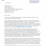
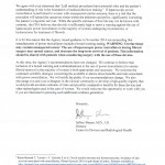
Jeffrey Shuren, M.D., J.D., the Director of the Center for Device and Radiological Health at the Food and Drug Administration, replied to an open letter from the Leiomyoma Morcellation Review Group.  You can read the original open letter here, and view Dr. Shuren’s reply in the images below.

from FDA 1from FDA 2

The Leiomyoma Morcellation Review Group sent the following letter to the FDA in response:

Dear Dr. Shuren,

Thank you for your response to our letter and for your invitation to continue the dialog regarding the FDA’s restriction on the use of the morcellator for the majority of women with uterine fibroids.  We respect the FDA’s difficult position in this matter, especially considering the adverse publicity and pressure  generated by Dr. Reed’s husband. As responsible physicians and women’s health-care advocates we think that women who need treatment for uterine fibroids, as with all patients, deserve to make treatment decisions based on the best information available.  We know that this is also something that the FDA believes as well.  We would like to make the following comments:

1) As we are sure you are aware, a recent publication from the Michigan Surgical Quality Collaboration showed that the rate of laparoscopic hysterectomies decreased and the rate of abdominal hysterectomies increased following the FDA’s restrictive policy on morcellation, and there was an associated concomitant increase in major surgical complications and 30-day readmissions to the hospital (Harris).  This increase in morbidity was anticipated by the decision analysis published by Siedhoff and colleagues.  Unintentionally, the FDA is compromising the health of some women.  Our concerns regarding higher surgical morbidity associated with increased use of laparotomy for women needing surgical treatment for fibroids drove our decision to prepare our letter questioning the FDA’s guidance regarding morcellation.  Unfortunately, our concerns are now validated by published data.

2) In our letter to the FDA and our commentary published in Obstetrics and Gynecology, we did “challenge” your methodology and calculation of the prevalence of leiomyosarcoma among women having surgery for presumed fibroids, as well as the impact of electro-mechanical morcellation on survival.  We are very concerned that your researcher did not carefully consider all of the published data available at that time.  We are also concerned that the FDA analysis was reported to be performed by a close friend of Dr. Noorchashm and that this conflict of interest was not disclosed during the hearing.

The Kaiser study that you cite is only one of many recent attempts to determine the prevalence of LMS among women having surgery for uterine fibroids.  In a further analysis, we added the women from the Bojahr study cited by us and the Raines-Bennet study cited by you and re-calculated our meta-analysis.  The rate of LMS then becomes 1:1924 or 0.052% and essentially no different from our original calculation.

3) In the Kaiser study you cite, our concern is that it appears that you incorrectly state that “the adjusted risk ratio for death at one year for power morcellated uteri was significantly greater compared to uteri that were not morcellated”.  In fact, there was no difference in one year survival in these groups of women, as noted in the abstract and indicated in Figure 3.    The only statistically significant finding in that analysis was the one-year comparison of women with any type of morcellation compared with no morcellation, suggesting that power morcellation is not the determining factor.  The analysis also showed no increased mortality at  2 or 3 years, consistent with an aggressive disease that has early spread by the hematogenous route.

Additionally, the FDA did not carefully considered the published literature. Indeed, the data used to determine that laparoscopic power morcellation “significantly worsening the patient’s long-term survival” has been criticized by us and others for being of poor quality characterized by heterogeneous referral populations, small numbers and suboptimal quantitative analysis.

Furthermore, since the vast majority of cases analyzed in the literature were morcellation procedures performed using a scalpel via either the vaginal or mini-laparotomy routes, it would seem reasonable to restrict of vaginal and mini-lap morcellation in addition to electro-mechanical morcellation.  Such a restriction would essentially limit surgical options for women with fibroids to total abdominal hysterectomy, a position that would be called into question by most women and their gynecologists.

We respect the concerns of the FDA staff.  However, the FDA’s  decision  to place a boxed warning, albeit intended to protect women and to make them aware of risks,  has in fact reduced treatment options for women, paradoxically increasing the risks associated with surgery for uterine fibroids. Although we are not questioning your intentions, your agency’s actions do not appear consistent with your mission to ensure the safety and health of all women.

4) While we agree that a registry might clarify the prevalence of LMS and the influence of morcellation on survival, the registry would need to be mandatory for all US women with uterine LMS.  With such a rare condition, anything less would be subject to significant selection and reporting bias.   We are aware that a registry of women with leiomyomas is currently being developed through funding from PCORI and that this will provide important prospective data.  It will be essential to have appropriate funding for an LMS registry as well.  We sincerely hope that the FDA is discussing this need with your sister federal agencies.

Women and their physicians look to the FDA for guidance on issues that impact public health and safety. We do not feel that women have been properly served by the FDA’s current morcellation advisory.  We request that the FDA reconsider the currently available data and re-evaluate the restrictions on the use of the electro-mechanical morcellator for the majority of women with uterine fibroids.  We welcome the opportunity to talk to you about these issues and would be willing to send a group to Washington for further discussion if you wish.  We are confident that the FDA shares with our group a common concern for the health and safety of all women with leiomyomas. Our goal is to find a way forward that will provide an appropriate and reasonable approach to uterine leiomyoma treatment and medical care.

Thank you for your consideration,

William H. Parker, MD, Santa Monica-UCLA Medical Center

Andrew M. Kaunitz, MD , University of Florida College of Medicine–Jacksonville

Elizabeth Pritts, MD, Middleton, WI

David Olive, MD, Middleton, WI

Eva Chalas, MD, FACOG, FACS, Winthrop-University Hospital, Mineola, NY

Daniel L. Clarke-Pearson, MD, UNC-Chapel Hill

Jonathan S Berek, MD, MMS, Stanford University School of Medicine

Barbara Goff, MD, University of Washington, Seattle, WA

Robert Bristow, MD, UC Irvine School of Medicine

Robin Farias-Eisner, MD, David Geffen School of Medicine at UCLA

Amanda Nickles Fader, MD, Johns Hopkins Medicine

G Larry Maxwell, MD, FACOG, COL(ret) U.S. Army, Inova Fairfax Hospital

Scott C Goodwin, MD, University of California, Irvine

Susan Love, MD, Dr. Susan Love Research Foundation, Los Angeles, California

William E Gibbons, MD, Baylor College of Medicine

Leland J. Foshag, M.D., FACS, John Wayne Cancer Institute, Santa Monica, California

Phyllis C. Leppert, MD, PhD, Duke University School of Medicine

Charles W. Nager, MD, UC San Diego Health System

Judy Norsigian, Our Bodies, Ourselves, Boston, Mass

Timothy Johnson, MD, University of Michigan

David S. Guzick, MD, PhD, University of Florida

Anton J. Bilchik, MD, PhD, FACS, John Wayne Cancer Institute, Santa Monica, California

Hugh Taylor, MD, Yale School of Medicine

Richard J. Paulson, MD, Keck School of Medicine, University of Southern California

Professor Cindy Farquhar, University of Auckland, NZ

Sawsan As-Sanie, MD MPH, University of Michigan

Linda D. Bradley, MD, Cleveland Clinic

Stacey A. Scheib, MD, Johns Hopkins Hospital

Carla Dionne, MPH, MBA, National Uterine Fibroid Foundation

Laurel W. Rice, MD , University of Wisconsin-Madison School of Medicine and Public Health

Alison Jacoby, MD, University of California, San Francisco

Charles Ascher-Walsh, MD, Mt. Sinai School of Medicine

G. David Adamson, MD, Stanford University School of Medicine

Matthew Siedhoff, MD MSCR, University of North Carolina at Chapel Hill

Robert Israel, M.D, Keck School of Medicine, University of Southern California

Marie Fidela Paraiso, MD, Cleveland Clinic

Michael M. Frumovitz, M. D., M.P.H., FACOG, FACS, The University of Texas MD Anderson Cancer Center

Guy I. Benrubi, M.D., University of Florida College of Medicine – Jacksonville

Steven S. Raman, MD, David Geffen School of Medicine at UCLA

Rosanne M Kho MD, Columbia University Medical Center

John R Lurain, MD, Northwestern University Feinberg School of Medicine

Ayman Al-Hendy  MD PhD FRCSC FACOG CCRP, Medical College of Georgia

Ted L. Anderson, M.D., Ph.D., Vanderbilt University School of Medicine

R. Kevin Reynolds, MD, FACS, FACOG, University of Michigan

John DeLancey, MD, University of Michigan Health System

View all blog archives

An open letter to the FDA regarding the use of morcellation procedures for women having surgery for presumed uterine fibroids

In November, 2014 the FDA ruled that power morcellation was contra-indicated in “the majority of women” having surgery for uterine fibroids due to the potential risk of spreading occult uterine sarcoma.  Although problems with this ruling were immediately apparent, the passage of time has allowed for more clarity on the related medical issues.

Prevalence of Leiomyosarcoma among women having surgery for presumed uterine fibroids

The prevalence of occult leiomyosarcoma among women with fibroids is critical for every patient.  All medical procedures have potential risk and the patient’s understanding of risk is the foundation of medical decision-making.

The FDA estimated that for every 458 women having surgery for fibroids, one woman would be found to have an occult leiomyosarcoma (LMS).  We challenge this calculation.  To estimate this risk, the FDA searched medical databases using the terms “uterine cancer” AND “hysterectomy or myomectomy”.   Because “uterine cancer” was required, studies where cancer was not found or discussed were not identified.  Nine studies, all but one of which were retrospective, were analyzed including a non-peer-reviewed letter to the editor and an abstract from an unpublished study. (Leung, Rowland)    Additionally,  three “leiomyosarcoma ” cases identified by the FDA do not meet current pathologic criteria for cancer and would now be classified as benign “atypical” leiomyomas.  If atypical leiomyomas and non-peer-reviewed data are excluded, the FDA identified 8 cases of LMS among 12,402 women having surgery for presumed leiomyomas, a prevalence of 1 in 1,550 (0.064%).

Pritts et al. recently published a more rigorous meta-analysis of 133 studies and determined that the prevalence of LMS among women having surgery for presumed fibroids was 1 in 1,960, or 0.051%.   All peer-reviewed reports in which surgery was performed for presumed fibroids were analyzed, including reports where cancer was not found. Inclusion criteria required that histopathology results be explicitly provided and available for interpretation. Among the 26 randomized control trials analyzed, 1,582 women had surgery for fibroids and none were found to have LMS.   Bojahr et al., recently published a large population-based prospective registry study and reported 2 occult LMS among 8,720 women having surgery for fibroids (0.023%).    In summary, the re-analyzed FDA dataset yields a prevalence of 1 in 1,550 (0.064%), the Pritts study reports a prevalence of 1 in 1,960 (0.051%) with the RCT’s having a prevalence of 0 and the Bojahr study reported a prevalence of 2 of 8,720 (0.023%). We acknowledge that with rare events statistical analysis may be uncertain and confidence Intervals may be wide.  However, these numbers do not support the FDA’s estimated prevalence of LMS among women having surgery for presumed fibroids and those at risk for morcellation of a leiomyosarcoma.

Prognosis for women with morcellated LMS

Leiomyosarcoma, removed intact without morcellation have a poor prognosis.  Based on SEER data, the 5 year survival of Stage I and II LMS is only 61%. (Kosary)   Whether morcellation influences the prognosis of women with LMS is not known and the biology of this tumor has not been well studied.  Distant metastasis occur early in the disease process, primarily hematogenous dissemination.  Four frequently quoted published studies examine survival following power-morcellation.  Surprisingly, virtually none of the women in these studies had power-morcellation.  Furthermore, the data presented in these reports are poorly analyzed and patient numbers are very small.  Park, et. al. reported only one of the 25 morcellated cases had laparoscopic surgery with power-morcellation.   Eighteen women had a laparoscopically-assisted vaginal hysterectomy with scalpel-morcellation performed through the vagina, one had a vaginal hysterectomy with scalpel-morcellation and 5 had mini-laparotomy with scalpel-morcellation through small lower abdominal incisions.  Seventeen of the 25 patients plotted in the published survival curve were referred to the hospital after initial diagnosis or the discovery of a recurrence at another institution. Since the number of non-referred women with less aggressive disease or without recurrence is not known, it is not possible to determine differences in survival between patients with and without morcellation.    In a study by Perri et. al., none of the patients had power-morcellation.   Four women had an abdominal myomectomy, four had a hysteroscopic myomectomy with tissue confined within the uterine cavity, two had a laparoscopic hysterectomy with scalpel-morcellation, four had a supra-cervical abdominal hysterectomy with cut-through at the cervix and two had an abdominal hysterectomy with injury to the uterus with a sharp instrument.   When comparing the outcomes for women with morcellated and non-morcellated LMS, Morice et. al., found no difference in recurrence rates or over-all and disease-free survival at six months.  In the only study to compare use of power- with scalpel-morcellation in women with LMS, Oduyebo et. al. found no difference in outcomes for the 10 women with power-morcellation and five with scalpel-morcellation followed for a median of 27 months (range, 2-93).    Notably, a life table analysis of the above studies showed no difference in survival between morcellation methods. (Pritts)

Of note, laparoscopic-aided morcellation allows the surgeon to inspect the pelvic and abdominal cavities and irrigate and remove tissue fragments under visual control. In contrast, the surgeon cannot visually inspect the peritoneal cavity during vaginal or mini-laparotomy procedures.  Morcellation within containment bags have recently been utilized in an attempt to avoid spread of tissue.  These methods have not yet been proven effective or safe, and there is  concern that bags may make morcellation more cumbersome and less safe.

What the FDA Restrictions Mean for Women

The FDA communication states, “the FDA is warning against the use of laparoscopic -morcellators in the majority of women undergoing myomectomy or hysterectomy for treatment of fibroids.”  This statement is not consistent with current evidence.  Moreover, a severe restriction of morcellation, including vaginal and mini-laparotomy morcellation, would limit women with symptomatic leiomyomas to one option, total abdominal hysterectomy. For women with fibroids larger than a 10-week pregnancy size, which most often require either scalpel or power-morcellation in order to remove tissue, a ban on morcellation would eliminate the following procedures:

  • vaginal hysterectomy (scalpel morcellation)
  • mini-laparotomy hysterectomy (scalpel morcellation)
  • laparoscopic hysterectomy (scalpel morcellation)
  • laparoscopic supra-cervical hysterectomy (cervix cut-through)
  • open supra-cervical hysterectomy (cervix cut-through)
  • laparoscopic myomectomy (power morcellation)
  • mini-laparotomy myomectomy (scalpel morcellation)
  • hysteroscopic myomectomy (intrauterine morcellation)
  • uterine artery embolization (no specimen and will delay diagnosis)
  • high-intensity focused ultrasound (no specimen and will delay diagnosis)

If abdominal hysterectomy is recommended to women with fibroids, will women be better off?

By focusing exclusively on the risk of LMS, the FDA failed to take into account other risks associated with surgery.   Laparoscopic surgery uses small incisions, is performed as an out-patient procedure (or overnight stay), has a faster recovery (2 weeks versus 4-6 for open surgery) and is associated with lower mortality and fewer complications.  These benefits of minimally invasive surgery are now well-established in gynecologic and general surgery.  Using published best-evidence data, a recent decision analysis showed that, comparing 100,000 women having laparoscopic hysterectomy with 100,000 having open hysterectomy, the group having laparoscopic surgery  would experience 20 fewer peri-operative deaths, 150 fewer women would have a pulmonary or venous embolus and 4,800 fewer women would have a wound infection. (Seidhoff)   Importantly, women having open surgery would have 8,000 fewer quality-of-life years.  A recently published study found that  in the eight months following the FDA safety communication, utilization of laparoscopic hysterectomies decreased by 4.1% (p=0.005) and both abdominal and vaginal hysterectomies increased (1.7%, p =0.112 and 2.4%, p=0.012, respectively). (Harris)  Major surgical complications (not including blood transfusions) significantly increased from 2.2% to 2.8% (p=0.015), and the rate of hospital readmission within 30 days also increased from 3.4% to 4.2% (p=0.025).  These observations merit consideration as women weigh the pros and cons of minimally-invasive surgery with morcellation versus open surgery.  These observations merit consideration as women weigh the pros and cons of minimally invasive surgery with possible morcellation vs. open surgery.

Clinical Recommendations

Recent attention to surgical options for women with uterine leiomyomas and the risk of an occult leiomyosarcoma is a positive development in that the gynecologic community is re-examining relevant issues. We respectfully suggest that the following clinical recommendations be considered:

  • The risk of LMS is higher in older post-menopausal women and greater caution should be exercised prior to recommending morcellation procedures for these women.
  • Preoperative consideration of LMS is important and women age 35 or older with irregular uterine bleeding and presumed fibroids should have an endometrial biopsy, which occasionally may detect LMS prior to surgery.  Women should have  normal results of cervical cancer screening.
  • Ultrasound or MRI findings of a large irregular vascular mass, often with irregular anechoic (cystic) areas reflecting necrosis, may cause suspicion of LMS.
  • Women wishing minimally-invasive procedures with morcellation, including scalpel-morcellation via the vagina or mini-laparotomy, or power-morcellation using laparoscopic guidance, should understand the potential risk of decreased survival should LMS be present.  Open procedures should be offered to all women who are considering minimally -invasive procedures for “fibroids”.
  • Following morcellation, careful inspection for tissue fragments should be undertaken and copious irrigation of the pelvic and abdominal cavities should be performed to minimize the risk of retained tissue.
  • Further investigations of a means to identify LMS pre-operatively should be supported.  Likewise, investigation into the biology of LMS should be funded to better understand the propensity of tissue fragments or cells to implant and grow.    With that knowledge, minimally- invasive procedures could be avoided for women with LMS and women choosing minimally-invasive surgery could be re-assured that they do not have LMS.

Respecting women who suffer from leiomyosarcoma, we conclude that the FDA directive was based on a misleading analysis.   Consequently, more accurate estimates regarding the prevalence of LMS among women having surgery for fibroids should be issued. Women have a right to self-determination.   Modification of the FDA’s current restrictive guidance regarding power-morcellation would empower each woman to consider the pertinent issues and have the freedom to undertake shared decision-making with her surgeon in order to select the procedure which is most appropriate for her.

William Parker, MD
Clinical Professor
UCLA School of Medicine
Director, Minimally Invasive Gynecologic Surgery
Santa Monica-UCLA Medical Center

Jonathan S Berek, MD, MMS
Laurie Kraus Lacob Professor
Director, Stanford Women’s Cancer Center
Director, Stanford Health Care Communication Program
Chair, Department of Obstetrics and Gynecology
Stanford University School of Medicine

Elizabeth Pritts, MD
Wisconsin Fertility Institute
Middleton, WI

David Olive, MD
Wisconsin Fertility Institute
Middleton, WI

Andrew M. Kaunitz, MD
University of Florida Research Foundation Professor
Associate Chair, Department of Obstetrics and Gynecology
University of Florida College of Medicine–Jacksonville

Eva Chalas, MD, FACOG, FACS
Chief, Division of Gynecologic Oncology
Director of Clinical Cancer Services
Vice-Chair, Department of Obstetrics and Gynecology
Winthrop-University Hospital

Daniel Clarke-Pearson, MD
Professor and Chair
Clinical Research, Gynecologic Oncology Program
UNC-Chapel Hill

Barbara Goff, MD
Professor of Obstetrics and Gynecology
Director, Division of Gynecologic Oncology
University of Washington
Seattle, WA

Robert Bristow, MD
Professor and Chair
Department of Obstetrics and Gynecology
UC Irvine School of Medicine

Hugh S. Taylor, M.D.
Anita O’Keeffe Young Professor and Chair Department of Obstetrics, Gynecology and Reproductive Sciences, Yale School of Medicine
Chief of Obstetrics and Gynecology
Yale-New Haven Hospital

Robin Farias-Eisner, MD
Chief of Gynecology and Gynecologic Oncology
Department of Obstetrics and Gynecology
David Geffen School of Medicine at UCLA

Amanda Nickles Fader, MD
Director of the Kelly Gynecologic Oncology Service
Director, F.J. Montz Fellowship in Gynecologic Oncology
Johns Hopkins Medicine

G Larry Maxwell, MD, FACOG, COL(ret) U.S. Army
Chairman, Department of Obstetrics and Gynecology, Inova Fairfax Hospital
Co-P.I., DOD Gynecologic Cancer Translational Research Center of Excellence
Professor, Virginia Commonwealth School of Medicine
Executive Director, Globe-athon to End Women’s Cancer

Scott C Goodwin, MD
Hasso Brothers Professor and Chairman, Radiological Sciences
University of California, Irvine

Susan Love, MD
Dr. Susan Love Research Foundation

William E Gibbons, MD
Professor
Director, Division of Reproductive Medicine
Director, Fellowship Training
Department of Obstetrics and Gynecology
Baylor College of Medicine
Chief, Reproductive Medicine the Pavilion For Women at Texas Children’s Hospital
Houston, Texas

Leland J. Foshag, M.D., FACS
Surgical Oncology, Melanoma and Sarcoma
John Wayne Cancer Institute
Santa Monica, California

Phyllis C. Leppert, MD, PhD
Emerita Professor of Obstetrics and Gynecology
Duke University School of Medicine
President, The Campion Fund, Phyllis and Mark Leppert Foundation for Fertility Research

Judy Norsigian
Co-founder, Our Bodies, Ourselves
Boston, Mass

Charles W. Nager, MD
Professor and Chairman
Department of Reproductive Medicine
UC San Diego Health System

Timothy Johnson, MD
Chair, Department of Obstetrics and Gynecology, University of Michigan
Arthur F. Thurnau Professor, Professor of Women’s Studies, and Research Professor in the Center for Human Growth and Development

David S. Guzick, MD, PhD
Senior Vice President, Health Affairs
President, UF Health
University of Florida

Sawsan As-Sanie, MD MPH
Assistant Professor
Director, Minimally Invasive Gynecologic Surgery and Fellowship
Director, Endometriosis Center
University of Michigan

Richard J. Paulson, MD
Alia Tutor Chair in Reproductive Medicine
Professor and Vice-Chair
Department of Obstetrics and Gynecology
Chief, Division of Reproductive Endocrinology and Infertility
Keck School of Medicine
University of Southern California

Professor Cindy Farquhar
Department of Obstetrics and Gynaecology and National Women’s Health
University of Auckland, NZ

Linda Bradley, MD
Vice Chair of Obstetrics, Gynecology and Women’s Health Institute
Director of The Fibroid and Menstrual Disorders Center
Cleveland Clinic
Dept of Obstetrics and Gynecology

Stacey A. Scheib, MD
Assistant Professor
Director of the Hopkins Multidisciplinary Fibroid Center
Director of Minimally Invasive Gynecologic Surgery
Johns Hopkins Hospital

Anton J. Bilchik, MD, PhD, FACS
Professor of Surgery
Chief of Medicine at John Wayne Cancer Institute
Santa Monica, California

Laurel W. Rice, MD
Chair of the Department of Obstetrics and Gynecology
Professor, Division of Gynecology Oncology
University of Wisconsin-Madison School of Medicine and Public Health

Carla Dionne
Founder, National Uterine Fibroid Foundation

Alison Jacoby, MD
Director, UCSF Comprehensive Fibroid Center
Interim Chief, Division of Gynecology
University of California, San Francisco

Charles Ascher-Walsh, MD
Director of Gynecology, Urogynecology, MIS
Mt. Sinai School of Medicine
New York, NY

Sarah J. Kilpatrick, MD, PhD
Chair, Department of Obstetrics and Gynecology
Associate Dean, Faculty Development
Helping Hand of Los Angeles Chair in Obstetrics and Gynecology
Cedars-Sinai Medical Center
Los Angeles, California

David Adamson, MD
Clinical Professor, Stanford University School of Medicine
Past President of the American Society for Reproductive Medicine

Matthew Siedhoff, MD MSCR
University of North Carolina at Chapel Hill
Obstetrics & Gynecology
Minimally Invasive Gynecologic Surgery, Director

Robert Israel, M.D
Professor, Department of OB/GYN
Chair, Quality Improvement
Director, Women’s Health Clinics and Referrals, LAC+USC Medical Center

Marie Fidela Paraiso, MD
Head, Female Pelvic Medicine & Reconstructive Surgery
Cleveland Clinic
Cleveland, OH

Michael M. Frumovitz, M. D., M.P.H., FACOG, FACS
Fellowship Program Director
Department of Gynecologic Oncology and Reproductive Medicine
The University of Texas MD Anderson Cancer Center

John R Lurain, MD
Marcia Stenn Professor of Gynecologic Oncology
Program director for the fellowship in gynecologic oncology
Northwestern University Feinberg School of Medicine

Ayman Al-Hendy  MD PhD FRCSC FACOG CCRP
GRU Director of Interdisciplinary Translational Research
MCG Assistant Dean for Global Translational Research
Professor and Director
Division of Translational Research
Department of Obstetrics and Gynecology
Medical College of Georgia
Georgia Regents University

Guy I. Benrubi, M.D.
Senior Associate Dean for Faculty Affairs,
Robert J. Thompson Professor and Chair,
Department of Obstetrics and Gynecology
University of Florida College of Medicine – Jacksonville

Steven S. Raman, MD
Professor of Radiology, Urology, and Surgery
Co-director of the Fibroid Treatment Program
David Geffen School of Medicine at UCLA

Rosanne M Kho MD
Associate Professor
Head, Section of Urogynecology and FPMRS
Co-Director, MIGS Fellowship Program
Columbia University Medical Center
New York, NY

Ted L. Anderson, M.D., Ph.D.
Betty and Lonnie S. Burnett Professor and Chair, Obstetrics and Gynecology
Division Director, Gynecology
Vanderbilt University School of Medicine

Kevin Reynolds, MD, FACS, FACOG
The George W. Morley Professor and Chief
Division of Gynecologic Oncology
University of Michigan
Ann Arbor Michigan

John DeLancey, MD
Norman F. Miller Professor of Obstetrics & Gynecology
University of Michigan Health System

All institutional affiliations are for identification purposes only

No signee has declared a conflict of interest


References

Bojahr B, De Wilde R, Tchartchian G.  Malignancy rate of 10,731 uteri morcellated during laparoscopic supracervical hysterectomy (LASH). Arch Gynecol Obstet 2015;292:665-672.

Kosary CL. SEER survival monograph: Cancer survival among adults: U.S. SEER program, 1988-2001, patient and tumor characteristics. In: Ries LAG, Young JL, Keel GE,

Eisner MP, Lin DY, Horner MD, eds. Cancer of the corpus uteri. Bethesda, MD: National Cancer Institute, SEER Program, NIH; 2007:123-32.

Harris JA, Swenson CW, Uppal S, Kamdar N, Mahnert N, As-Sanie S, Morgan DM. Practice Patterns and Postoperative Complications Before and After Food and Drug Administration Safety Communication on Power Morcellation. Am J Obstet Gynecol (2015), doi:10.1016/j.ajog.2015.08.047.

Leung F, Terzibachian J. Re: “The impact of tumor morcellation during surgery on the prognosis of patients with apparently early uterine leiomyosarcoma [letter]. Gynecol Oncol 2012;124:172-3

Lieng M, Berner E, Busund B. Risk of morcellation of uterine leiomyosarcomas in laparoscopic supracervical hysterectomy and laparoscopic myomectomy, a retrospective trial including 4791 women. J Minim Invasive Gynecol. 2015;22:410-4

Morice P, Rodriguez A, Rey A, Pautier P, Atallah D, Genestie C, Pomel C, Lhommé C, Haie-Meder C, Duvillard P, Castaigne D. Prognostic value of initial surgical procedure for patients with uterine sarcoma: analysis of 123 patients. Eur J Gynaecol Oncol. 2003;24:237-40.

Oduyebo T, Rauh-Hain AJ, Meserve EE, Seidman MA, Hinchcliff E, George S, Quade B, Nucci MR, Del Carmen MG, Muto MG. The value of re-exploration in patients with inadvertently morcellated uterine sarcoma. Gynecol Oncol. 2014;132:360-5.

Park JY, Park SK, Kim DY, Kim JH, Kim YM, Kim YT, Nam JH. The impact of tumor morcellation during surgery on the prognosis of patients with apparently early uterine leiomyosarcoma. Gynecol Oncol. 2011;122:255-9.

Perri T, Korach J, Sadetzki S, Oberman B, Fridman E, Ben-Baruch G. Uterine leiomyosarcoma: does the primary surgical procedure matter? Int J Gynecol Cancer. 2009;19:257-60

Pritts E, Vanness D, Berek  J, Parker W, Feinberg R, Feinberg J, Olive D. The prevalence of occult leiomyosarcoma at surgery for presumed uterine fibroids: a meta-analysis. Gynecol Surg. accessed on-line 5/30/15 at: http://link.springer.com/article/10.1007/s10397-015-0894-4

Pritts E, Parker W, Brown J, Olive D. Outcome of occult uterine leiomyosarcoma after surgery for presumed uterine fibroids: a systematic review. J Minim Invasive Gynecol. 2015;22:26-33

Rowland M, Lesnock J, Edwards R, Richard S, Zorn K, Sukumvanich P, et al. Occult uterine cancer in patients undergoing laparoscopic hysterectomy with morcellation (abstract) Gynecol Oncol 2012;127:S29

Siedhoff MT, Wheeler SB, Rutstein SE, Geller EJ, Doll KM, Wu JM, Clarke-Pearson DL. Laparoscopic hysterectomy with morcellation vs abdominal hysterectomy for presumed fibroid tumors in premenopausal women: a decision analysis.  J Obstet Gynecol. 2015;212:591.e1-8

View all blog archives

Efficacy and safety of repeated use of ulipristal acetate in uterine fibroids

Authors: Donnez J, and others

Journal: Fertility & Sterility. 2015 Feb;103:519-27

Study From: Brussels, Belgium and 12 other European medical centers

Problem:  Ulipristal, currently available in Europe and Canada, is an oral medication that blocks the action of progesterone on fibroids and stops heavy bleeding and decreases the size of fibroids. In an earlier study of this medication, it was shown to be effective for 13 weeks of treatment without worrisome side-effects.  The current study examined whether the medication was still safe if women were given 3 months of treatment followed by 2 months off the medication and then another 3 months of treatment.

Study:  451 women with symptomatic uterine fibroids and heavy bleeding were given 3 months of treatment, followed by 2 months off the medication, followed by another 3 months of treatment.

Results: About 70% of women had no bleeding while on the medication and women did resume periods (but, lighter bleeding than before any treatment) during the two months they were off of the medication.  After the second treatment, fibroids had decreased in size by about 50%. Less than 5% of patients stopped the medication due to side-effects.  Pain and quality-of-life improved for most women.

Authors’ Conclusions:  Repeated 3 month treatments with daily oral ulipristal effectively controlled bleeding and pain, reduced fibroid volume, and restored quality-of-life in women with symptomatic fibroids.

Dr. Parker’s Comments:

Ulipristal appears to be a promising medication for the treatment of the symptoms often associated with fibroids: heavy bleeding and pelvic/abdominal pressure or pain.  An earlier study found the medication was safe for 13 weeks and the current study shows it is safe and effective when given 3 months on, two months off and three months on again.

In a normal menstrual cycle, the ovaries make estrogen in the beginning of the cycle which stimulates the uterine lining cells to grow. When ovulation occurs in the middle of the cycle, the ovary starts to produce progesterone in addition to the estrogen.  Progesterone makes the lining cells stop growing and gets the cells ready for implantation of a fertilized egg.  Up to now, most medications that block the action of progesterone have been shown to cause an overgrowth of the uterine lining cells because there is no brake on the cells’ growth.  The fear has been that this overgrowth could lead to cancer (a severe overgrowth) of the uterine lining cells.   However, with the two months off medication in between the treatments, this does not appear to happen.

My sense is that this medication will be an excellent treatment option for women who are close to menopause.  Once menopause starts, the ovaries stop making estrogen and progesterone so that all bleeding stops and fibroids usually shrink.  It remains to be seen whether long treatment (many months or years) will continue to be safe, but this medication does look very promising.  This medication is not yet available in the US, but is available in Europe, Canada and many other countries.

View all blog archives

Dr. Parker’s Discharge Instructions and Suggestions after Open Abdominal Surgery

UCLA Gyn Subspecialties Group
1450 Tenth Street, Suite 404
Santa Monica, CA 90401
(310) 451 8144 (phone)

Disclaimer: it is important for you to follow your own doctor’s instructions.

HOSPITAL RECOVERY

In the hospital, the nurses will remove the bladder catheter a few hours after surgery and they will ask you to sit in a chair and walk to the bathroom the night of surgery.  You will have the ON-Q pain pump (see below), but if you need other pain medication, ask the nurses and they will give it to you.  By the morning after surgery, you will be eating regular food and you will be able to walk in the hall.  Recent studies of this “enhanced surgical recovery” show that a quicker return to normal activities and diet was associated with faster patient recovery and a shorter hospital stay.

ACTIVITY AFTER SURGERY:

  • No strenuous exercise or intercourse for 4-6 weeks
  • You may start walking for exercise on day 1
    • Walking is the best activity following surgery.  You can walk up and down stairs, outside, and on a treadmill.  Walking will keep your circulation going and build back your stamina.
  • Fatigue: fatigue is extremely common after surgery and may last many weeks.
  • Iron – to help build back any blood lost during surgery. Either Fergon or SlowFe – 1-3 times a day along with 1,000mg of vitamin C (helps with the absorption of iron)
  • You may start driving when you are no longer taking narcotic pain medication and you feel it is SAFE for you to drive.
  • Showers only (no baths) for the first 2 weeks

PAIN/DISCOMFORT MANAGEMENT

  • For pain, we will prescribe either Norco 5/320mg or Percocet 5/325mg for you to take once you get home.
  • Days 2-3 may be worst, pain-wise
  • Some cramping and shooting pains are normal, even in areas away from the incisions
  • Do not drive under the influence of narcotic pain medication
  • “ON-Q” pain pump: I use an innovative device to relive post-operative pain called the ON-Q pain pump. As we are closing the incision, two tiny plastic tubes are inserted underneath the incision. The tubes are connected to a tennis ball sized pump that holds a solution of local anesthesia.  The pump slowly and automatically pushes the local anesthetic into the incision – exactly where you need it You don’t need to touch it). The ON-Q pump greatly reduces the need for injected narcotic pain medication doesn’t cause the grogginess that often accompanies narcotics.  The device lasts 3-4 days and then is easily, and absolutely painlessly, removed in the office when the suture is removed.
  • Lowering, raising and turning yourself will hurt (try using your arms instead of abs)
  • Urination may burn/pinch initially, but will get easier (avoiding caffeine). Taking D-Mannose helps.

PERIODS

  • Some bleeding and/or clumpy discharge is normal for a few weeks.
  • Periods won’t be normal again for 3-4 cycles

CALL US AT 310-451-8144 FOR:

Fever over 100.4 degrees F, bad pain unrelieved by pain medications, heavy bleeding, vomiting, dizziness, pus-like drainage from the wound or other concerns.

 DIET AND BOWEL ISSUES

  • No dietary restrictions
    • Avoid foods that cause gas such as broccoli, cauliflower and brussel sprouts
  • No alcohol if taking narcotic pain medications
  • Chewing gum helps with bowel function
  • Walking helps bowel function
  • Constipation is normal for a few days after abdominal surgery.  You may take Metamucil (fiber) or colace as needed.
  • Expect some gas discomfort/pain.  Gas pain can be relieved by walking, taking medication (Mylicon, Gas-X) and chewing gum.  Bending your legs while lying on your back may also help.
  • Bloating (“swelly belly”) is normal for up to 6-8 weeks and will go away by itself.  Surgery irritates and slows down the intestines and they retain more gas and stool. Some women find that an abdominal binder reduces pressure on the incision and helps them feel more support.  You can buy an abdominal binder at a surgical supply store or pharmacy.

INCISION HEALING

  • Skin incision – I close the skin incision with a suture technique that I learned from a plastic surgeon.  I use one continuous suture that is removed about 5-7 days after surgery, so that no suture material is left under the skin.  This technique decreases the chance of a thick scar.  I then place steri-strips (like band-aids) across the incision to take the tension off of the skin, which also decreases the risk of a thick scar.  The suture is quickly and painlessly removed in the office by our nurses and, if needed, the steri-strips are   replaced.  There are many layers of suture below the skin holding the incision together and these sutures dissolve a few months after surgery.
  • It is OK to shower with steri-strips (do not scrub the wound). You can remove the steri-strips after a week. After the steri-strips are removed, I suggest you buy Scar Away at the drug store.  Scar away is a silicon bandage (like a large band-aid) and you keep it on the incision for as many hours a day as you can.  Over time, the scar away thins and lightens the incision so that it heals more nicely.
  • Bruising: bruising around the incision(s) is very common, even beyond the surgery area, for a few weeks after surgery.  Cold packs can keep the incision swelling down and the bruising will go away by itself.
  • Incision “Shelf”: the first scar tissue that forms in the incision is made up of a thick, stiff collagen, which pulls the scar inward and leads to a “shelf” over the incision.  Over the next 3-6 months, the first collagen is slowly replaced by a more elastic collagen and the incision becomes flat with the rest of the skin.
  • Numbness: when we make the initial incisions to get to the uterus, we stretch, tear or even cut the very small nerves that go to the skin on the abdomen.  As a result, it is very common to have numbness, tingling, burning or itching above the incision for a few months after surgery.

APPOINTMENTS AFTER SURGERY:

  • Suture and ON-Q pain pump removal 5-7 days after surgery – with Dr. Parker’s nurse or nurse practitioner
  • Appointment with Dr. Parker 2 weeks after surgery
  • Appointment with Dr. Parker 6 weeks after surgery

SUGGESTED TO WEAR/BRING TO THE HOSPITAL

  • Comfortable slip-on flat shoes
  • Pants or skirt with elastic or drawstring waistband
  • Pillow for the car ride home (leave in car to put between your stomach and seat belt)
  • Lip balm (your mouth may be parched)
  • Your ID
  • Advance directives (optional)

BEFORE SURGERY –  SUGGESTED SHOPPING LIST

  • Throat lozenges: Sometimes the tube placed down your windpipe (trachea) can cause irritation which can feel like a sore throat.   At times, this irritation can lead to a not very pleasant feeling cough.  Lozenges can help soothe your throat and also help minimize the coughing.
  • Stool softener: Many types are available:  Smooth Move tea, Colace, Peri-Colace (stool softener and mild laxative together).  High-fiber foods.
  • Anti-gas pains: Mylicon, Maalox, Gas-X or peppermint tea
  • Three days of groceries/easy-prep foods, since you won’t feel like cooking. Consider broth/soup, fruits + veggies (avoid gassy ones like broccoli, cauliflower and Brussels sprouts), rice, crackers, juice, yogurt.

BEFORE MYOMECTOMY

We ask women to insert 2 tablets (Cytotec) vaginally about  2 hours before surgery. Cytotec has been shown to decrease blood loss during myomectomy surgery.  However, it can also cause cramping, vaginal bleeding and, sometimes nausea.   If you have any of these symptoms, they are normal (and we apologize), but the medication does help decrease surgical blood loss.

View all blog archives

Share/Save/Bookmark

Disclaimer: The ideas, procedures and suggestions contained on this web site are not intended as a substitute for consulting with your physician. All matters regarding your health require medical supervision.

Fibroid Doctor William H. Parker

Dr. William H. Parker is a board-certified Fellow in the American College of Obstetricians and Gynecologists. Dr. Parker is an internationally recognized expert in fibroid surgery and research. Based in San Diego, California, he is considered one of the best fibroid surgeons for abdominal and laparoscopic myomectomy in the United States and abroad. He has been chosen for Best Doctors in America and Top Doctors every year beginning in the late 90's.

Free Email Updates

Powered by Google FeedBurner : Receive free updates right to your inbox every time new content is added to this website.


Subscribe
Schedule an Appointment Today!
Read Dr. Parker's Blog

Blog Categories